Call today:
New Patients Call:
VISIT OUR OTHER LOCATIONS
Digital X-Ray is a form of X-ray imaging where digital X-ray sensors are used instead of traditional photographic film.
Some advantages of digital X-rays include time efficiency through bypassing chemical processing and the ability to digitally transfer and enhance images.
Also, less radiation can be used to produce an image of similar contrast to conventional radiography.
Instead of X-ray film, digital radiography uses a digital image capture device. This provides advantages like immediate image preview and availability, elimination of costly film processing steps, a wider dynamic range (which makes it more forgiving for over- and under-exposure), as well as the ability to apply special image processing techniques that enhance overall display quality of the image.
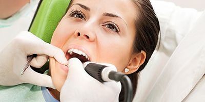
One of the intraoral camera’s most important advantages is the ability to capture and manipulate an image in the oral cavity and save it to the patient’s chart. If electronic patient charts are not being used, the images can be printed and filed in the patient’s paper chart. This allows dentists to share their observations with patients so they can become better informed about their own overall dental health.
The images captured by intraoral cameras are used to accurately visualize problems with teeth and/or tissues, including fractured teeth or restorations, various lesions, plaque buildup, and bleeding or inflamed tissues. With the intraoral camera’s technology, our dentists and their patients can collaborate on the right treatment plan to achieve optimal oral and dental health. Uxbridge Family Dental patients are now able to view potential problems along with our doctors while their treatment progress is effectively monitored. Sequential photos of the treatment effectively documents our patient compliance and improvement in their oral health.
With the use of the intraoral camera, our doctors can examine the oral cavity for any precautionary symptoms that may indicate a health risk. This includes, but is not limited to, images of any problematic oral lesions, eating disorders, and other diseases that may be present in the oral cavity that are essential to effective patient documentation.
The intraoral images captured by the camera closely illustrate the diagnoses and allow dentists to explain further to patients how to proceed with oral health treatment. This also allows dental care/oral health care providers to effectively demonstrate consequences to patients if the condition is left untreated.
Intraoral images can be printed out for patients to take home. Patients can then take time to consider the proposed treatment plan and hopefully become more motivated to improve their oral health.
Some intraoral software programs allow images to be modified in real time to illustrate proposed esthetic changes prior to altering the tooth structure. Additionally, images can be sent electronically to dental laboratories for a more accurate color match in veneers, crowns, and/or fixed partial dentures.
Digital X-Ray is a form of X-ray imaging where digital X-ray sensors are used instead of traditional photographic film.
Some advantages of digital X-rays include time efficiency through bypassing chemical processing and the ability to digitally transfer and enhance images.
Also, less radiation can be used to produce an image of similar contrast to conventional radiography.
Instead of X-ray film, digital radiography uses a digital image capture device. This provides advantages like immediate image preview and availability, elimination of costly film processing steps, a wider dynamic range (which makes it more forgiving for over- and under-exposure), as well as the ability to apply special image processing techniques that enhance overall display quality of the image.